Lengthening of Amyloid Fibrils May Lead to Nerve Cell Loss in FAP Patients, Study Suggests


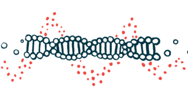
The distortion and subsequent atrophy of cells in the peripheral nervous system, due to the accumulation and lengthening of amyloid fibrils, may be behind nerve cell loss in patients with familial amyloid polyneuropathy (FAP), a study reports.
Researchers also identified dot-like structures outside cells that looked like the precursors of amyloid fibrils, or aggregates. As these structures assemble and enlarge into fibrils, they may pull surrounding cells, eventually causing damage to neurons.
The study, “The morphology of amyloid fibrils and their impact on tissue damage in hereditary transthyretin amyloidosis: An ultrastructural study,” was published in the Journal of Neurological Sciences.
FAP, also known as hereditary transthyretin amyloidosis, is a rare condition marked by abnormal deposits of a protein called transthyretin in the body’s organs and tissues, particularly in the nerves and the heart, leading to their progressive damage and eventually failure.
The condition is caused by mutations in the TTR gene that are thought to change the structure of the protein, preventing it from forming a functional shape. Rather, the defective protein gathers in deposits known as amyloid fibrils, which accumulate inside tissues.
However, how these fibrils lead to tissue damage is not yet fully understood.
To gather more clues, researchers investigated the shape of amyloid fibrils and their effect on neighboring tissues in the nerve tissue of patients with FAP.
They looked at biopsy or autopsy samples from 34 patients under an electron microscope, a type of microscope that uses electrons to zoom in and can obtain extremely high magnifications.
Scientists observed that the shape of amyloid fibrils varied between patients. Long fibrils were abundant in patients with early-onset Val30Met (the most common FAP mutation) FAP, whereas shorter ones marked late-onset Val30Met and non-Val30Met cases.
In addition, the amyloid filaments seemed to mature from dotty structures outside cells and pull the surrounding tissues during the maturation process.
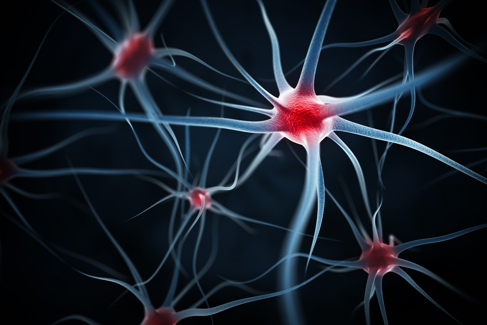
These fibrils, especially the long ones, considerably distorted neighboring cells, in particular Schwann cells. These cells wrap around neurons, providing them with support and covering them with myelin — a fat-rich protective layer that is key for the efficient conduction of impulses between neurons.
In patients’ tissues, the degeneration of Schwann cells was evident, particularly in those completely surrounded by amyloid fibrils. In contrast, nerve fibers that conserved myelin and whose shape was preserved tended to be only partially surrounded by the amyloid filaments. Researchers attribute this to the fact that nerve fibers are larger than Schwann cells, and less prone to become crooked due to amyloid fibrils.
Based on their results, the team concluded that “distortion and subsequent atrophy of Schwann cells may relate to nerve fiber loss” in FAP patients.
“We hypothesize that the traction of surrounding tissues by amyloid fibrils becomes more conspicuous as the fibrils become larger. In addition to the direct toxicity of amyloid fibrils, this mechanical distortion of surrounding tissues … may participate in the tissue damage, particularly in smaller diameter nerve fibers,” they said.