Magnetic Resonance Imaging (MRI) in FAP: What Does It Involve?

If your doctor suspects you have familial amyloid polyneuropathy (FAP), you will be asked to undergo diagnostic tests to confirm the presence of amyloid deposits in your body. Doctors then often use magnetic resonance imaging (MRI) to determine the deposits’ effect on your nervous system, heart, and other organs.
Here is some information about an MRI and what to expect.
What is FAP?
FAP is an inherited, progressive disorder characterized by the abnormal deposits of proteins called amyloids around peripheral nerves and other tissues. The buildup of amyloids leads to a host of symptoms. Heart enlargement and irregular heartbeats are the most serious.
How do doctors diagnose it?
Accumulation of amyloid deposits in the heart can cause the heart walls to thicken. This may result in enlarged heart muscles, and symptoms that include shortness of breath, fatigue, poor exercise capacity, and chest pain. Amyloid deposits can also interfere with the electrical impulses of the heart, leading to an irregular heartbeat and heart block.
Physicians who suspect heart involvement may order imaging tests to confirm a diagnosis, visualize the extent of heart damage, and plan for proper treatment. To evaluate heart involvement, non-invasive imaging techniques such as MRI are usually required.
What is MRI?
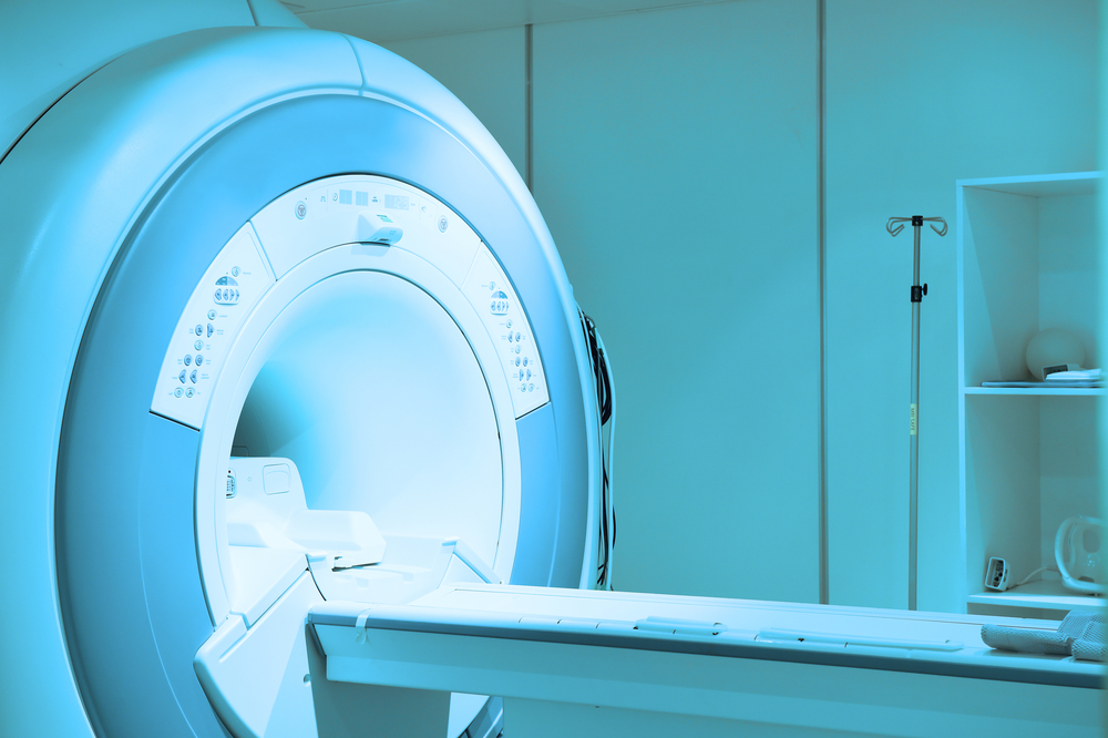
MRI is a scanning technology to produce three-dimensional and detailed images of the body. It uses strong magnetic fields and radio waves, but not ionizing radiation or X-rays.
Doctors often use MRIs to detect or diagnose disease, and to monitor the effect of treatment.
What to do before the MRI?
MRI may not be recommended in certain situations, including for people with a fitted metal implant such as a pacemaker or artificial joint. You also need to alert the radiology staff if you are pregnant, have a history of kidney problems or diabetes, have cochlear implants, aneurysm clips, skin tattoos, shrapnel or bullet wounds, artificial heart valves, or an implanted drug infusion device.
Leave jewelry and other valuables at home before undergoing an MRI scan, and bring a list of your current medications. Let your doctor know if you have anxiety-related claustrophobia, as you may be prescribed an oral medication to take for your MRI appointment. The scan itself is a painless and safe procedure, and typically manageable with a radiographer’s support.
Arrive at least a half-hour before your examination to fill out paperwork, and to change into a hospital gown.
What will happen during the MRI?
The radiographer (a person trained in imaging) will ask you to lie flat on a bed that will be moved into a tube called an MRI scanner, which contains powerful magnets. He or she will talk to you through an intercom, and watch you on a TV monitor throughout the scan.
You will be given earplugs or headphones to wear during the scan to muffle tapping its noises. There will also be an alarm button to alert the radiologist if you experience discomfort during the procedure.
You will need to lie still throughout the scan. You may also need to hold your breath for up to 30 seconds when requested. A scan usually lasts between 15 and 90 minutes, depending on the area being examined and number of images taken.
What happens after the MRI?
The radiologist will review the images and report their findings to your physician, who will meet with you and discuss further steps.
Last updated: Oct. 1, 2020
***
FAP News Today is strictly a news and information website about the disease. It does not provide medical advice, diagnosis, or treatment. This content is not intended to be a substitute for professional medical advice, diagnosis, or treatment. Always seek the advice of your physician or other qualified health providers with any questions you may have regarding a medical condition. Never disregard professional medical advice or delay in seeking it because of something you have read on this website.


