Nuclear Medicine Imaging Can be Used to Distinguish Amyloidosis Subtypes, Study Says


A nuclear imaging technique called 99mTc-DPD scintigraphy can be used to distinguish different subtypes of amyloidosis, including familial amyloid polyneuropathy (FAP), according to a new study.
The study, “99mTc-DPD scintigraphy and SPECT/CT in patients with AL and ATTR type amyloidosis: Potential clinical implications,” was published in the journal Medicine.
Amyloidosis refers to a group of diseases characterized by the buildup of protein aggregates, or clumps, in certain tissues or organs, damaging and impairing their normal function. There are many subtypes of amyloidosis, depending on the particular protein that starts to form aggregates, its distribution in the body, and the extent of these deposits.
In amyloid light-chain (AL) amyloidosis, the disease is triggered by the accumulation of small pieces of antibodies called light-chains, which are produced excessively by certain types of immune cells.
In contrast, in transthyretin (ATTR) amyloidosis, the disease is associated with the buildup of a protein called transthyretin. This may happen due to gene mutations in the TTR gene that provides instructions to make transthyretin — in the case of hereditary ATTR, or FAP — or in the absence of genetic alterations, known as wild-type ATTR.
Accurately characterizing the type of amyloidosis an individual has is essential, since the disease’s clinical course, treatment, and prognosis vary by subtype. Generally, a tissue biopsy is conducted to distinguish between the different subtypes of the disease. However, these procedures tend to be highly invasive.
For that reason, there is a need for a non-invasive imaging technique for amyloidosis that can screen the whole body. That would enable physicians to identify the particular type of amyloidosis, assess disease severity, and monitor a patient’s response to treatment.
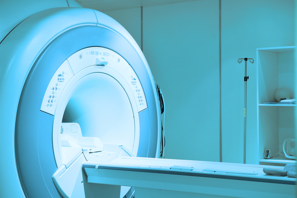
Imaging techniques based on nuclear medicine use small amounts of radioactive materials, known as radiotracers, that are injected into the bloodstream, inhaled or swallowed. Once in the body, these radiotracers are taken up by tissues, allowing physicians to obtain high-detailed images of the entire body in a single session.
“However, studies showing disease extent throughout the whole body using scintigraphy [nuclear medicine imaging] are scarce, even though amyloidosis is a systemic disease,” the researchers said.
To address this issue, a team of Korean researchers investigated the utility of 99mTc-DPD scintigraphy — a type of nuclear medicine imaging that uses a technetium-labeled radioactive compound (99mTc-DPD) – in 16 patients with systemic AL amyloidosis and five with ATTR amyloidosis.
Previous clinical findings from the 21 patients enrolled in the study revealed they had signs of the disease in 41 organs — 29 organs among those with AL amyloidosis and 12 organs among those with ATTR amyloidosis.
The imaging findings showed a significant 99mTc-DPD uptake in 24 of the 29 affected organs among those with AL amyloidosis, which led to a sensitivity — the ability of a test to correctly identify those with a disease — of 82.8%.
Among those with ATTR, a significant 99mTc-DPD uptake was observed in nine of the 12 affected organs, corresponding to a sensitivity of 75.0%.
When investigators compared the two disease subtypes with regard to 99mTc-DPD organ uptake, most patients (59.3%) with AL amyloidosis had grade 1 organ involvement. Grade 1 is organ uptake lower than that of the spine.
In contrast, nearly half of the individuals (41.7%) with ATTR amyloidosis had a grade 3 organ involvement — organ uptake greater than that of the spine. Hence, the degree of 99mTc-DPD organ uptake was significantly higher in people with ATTR compared with those with AL amyloidosis.
For 20 of the 41 affected organs, additional single photon emission computed tomography (SPECT) or computed tomography (CT) scan images were helpful in determining the presence of abnormal soft tissues.
The researchers also found the uptake of the radiotracer was higher in the heart of patients with ATTR amyloidosis than in those with AL amyloidosis, which was consistent with previous studies. Additionally, 99mTc-DPD scintigraphy demonstrated higher uptake in soft tissues among those with ATTR amyloidosis, which was not observed in patients with AL amyloidosis.
These findings indicate “that 99mTc-DPD scintigraphy might have capacity to differentiate between AL and ATTR subtypes with good sensitivity in various organs involving primary systemic AL and ATTR amyloidosis,” they concluded.