Blood Biomarkers Show Inflammation in FAP Patients Before Amyloid Deposits Evident, Study Find


Changes in the immune system and in inflammation may occur before the formation and accumulation of amyloid deposits in people with familial amyloid polyneuropathy, with biomarkers of these alterations easily evident in the blood, new research finds.
The study, “Inflammatory profiling of patients with familial amyloid polyneuropathy,” were published in BMC Neurology.
Familial amyloid polyneuropathy (FAP), also known as amyloid TTR variant (ATTRv), is a rare progressive disease caused by genetic mutations in the TTR gene, which provides instructions for making a protein called transthyretin.
When mutations occur, the structure of transthyretin changes, preventing it from binding to other transthyretin proteins, a process necessary for its normal transport function. The abnormal TTR protein then starts to form amyloid deposits that accumulate in tissues — including nerves, the heart, kidneys and eyes — slowly causing damage and eventually giving rise to symptoms associated with FAP.
“The diagnosis of ATTRv is challenging, often relying on genetic tools to identify TTR mutations as well as on the identification of Congo Red-positive amyloid deposits in biopsies usually taken from sural nerve [a nerve in the leg] and salivary glands,” the investigators wrote. But, they added, such symptoms are typically “absent” in those “asymptomatic or early symptomatic, supporting the need for novel biomarkers to identify patients in earlier disease phases.”
Previous studies have “indicated the possibility that inflammation may play a role in ATTRv pathogenesis” or disease development, the study continued, but “there are no data available showing which specific inflammatory components are altered in humans.”
Looking for new biomarkers that might identify patients at early stages of FAP, a group of researchers with the Universidade Federal do Rio de Janeiro in Brazil analyzed the serum of 28 ATTRv patients and 24 age- and sex-matched healthy individuals (controls) to look for markers of inflammation.
Among the 28 ATTRv patients enrolled, six were asymptomatic, 16 had mild sensory neuropathy in the lower limbs and could walk unassisted, and six had moderate-to-severe neuropathy in all limbs and required assistance to move or relied on a wheelchair.
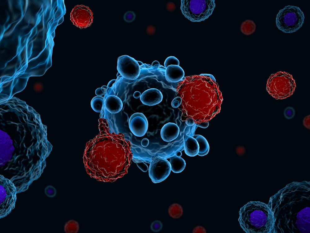
Results showed that serum levels of six out of the nine cytokines (molecules that mediate the immune and inflammatory response) analyzed — TNF-α, IL-1β, IL-8, IL-33, IFN-β and IL-10 — were significantly higher in ATTRv patients compared to healthy controls.
The serum levels of the remaining three cytokines studied — IL-12, IL-6 and cortisol — were either unchanged (IL-6 and cortisol) or significantly lower in ATTRv patients compared to controls (IL-12).
Interestingly, researchers found the serum levels of some of these cytokines (IL-33, IL-1β and IL-10) were abnormally high in asymptomatic patients, indicating that inflammation may be present well before amyloid deposits begin to accumulate in different tissues.
Analyses also showed that TNF-α levels directly correlated with disease progression, suggesting this particular cytokine may be one of the underlying mechanisms driving ATTRv development.
“The novel observation of alterations in immune response in serum from ATTRv patients suggests an important role of inflammation in ATTRv pathogenesis,” the investigators wrote. “This new focus on inflammation in ATTRv pathogenesis and progression provides new directions towards understanding this fatal disease and improving clinical criteria for choosing ATTRv patients that should receive tafamidis [Vyndaqel and Vyndamax; by Pfizer] treatment.”
“The drug has already showed positive results in clinical trials, but due to current clinical criteria is being administered to ATTRv patients that are already debilitated by disease,” they added.
“If these inflammatory markers could be evaluated in other ATTRv populations showing a similar profile with disease progression, we envision the possibility of using them as ATTRv biomarkers, which could be assessed vary rapidly and very easily.”