Imaging Is as Good as Tissue Sampling at Detecting Amyloid Deposits in Some Organs, Study Finds


Body-wide imaging can detect amyloid protein deposits in the organs of people with familial amyloid polyneuropathy (FAP), making it possible to assess the effects of new therapies without performing invasive tissue biopsies, a Japanese study reports.
Imaging can also be used to see whether drugs that remove amyloid build-ups from organs can alleviate the disease’s symptoms as well, researchers said.
A shortcoming is that imaging fails to detect amyloid build-ups in nerve structures. Both FAP and another condition the team studied, systemic immunoglobulin light-chain (AL) amyloidosis, are neurodegenerative diseases.
The study, “Visualization of multiple organ amyloid involvement in systemic amyloidosis using 11C-PiB PET imaging,” appeared in the European Journal of Nuclear Medicine and Molecular Imaging.
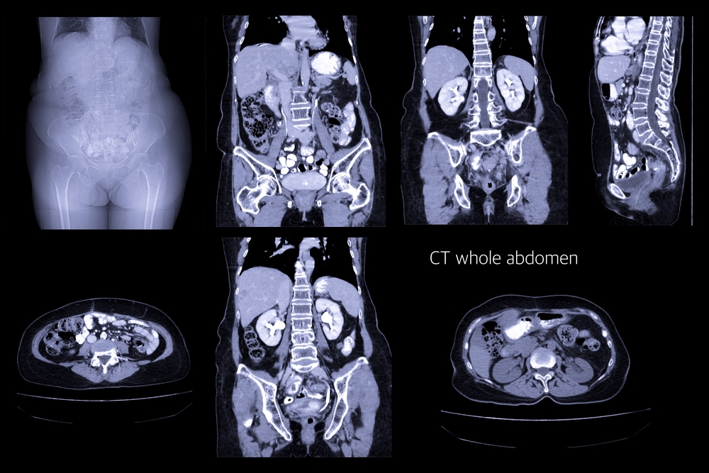
Researchers at Japan’s Shinshu University School of Medicine used an imaging technique called positron emission tomography. PET scans work by emitting a radioactive tracer molecule that binds to a target molecule.
To see whether PET scans could detect amyloid deposits, the researchers recruited seven people with FAP, a disease that is also called hereditary transthyretin amyloidosis, or TTR amyloidosis. The team also examined seven people with AL and three healthy controls.
They discovered that the imaging detected high levels of amyloid in the heart and stomach, which correlated with FAP and AL’s manifestations.
Unfortunately, PET scans also detected deposits in tissue that was not generating symptoms of disease, such as the tongue.
In addition, imaging did not detect amyloid deposits in the peripheral nervous system — the parts of the system outside the brain and spinal cord. This was despite the fact that six FAP patients and one AL patient were experiencing obvious nerve malfunctioning.
Researchers also found amyloid deposits in some FAP patients’ lachrymal gland, brain, scalp, nasal mucosa, and throat. The lachrymal glands generate the tears that moisten and cleanse the eyes.
As an additional test, researchers compared the scan results with the amount of amyloid in tissue samples. They concluded that imaging identified amyloid deposits as examining tissue with a microscope.
While imaging can shed light on the severity of a neurodegenerative disease, particularly in assessing new treatments, researchers admitted that its inability to detect amyloid deposits in nerve structures was a shortcoming.